The bar patterns are tiny images of a resolution chart, formed by the ommatidial lenses in the compound eye of a shed cuticle of a Brown Marmorated Stink Bug.
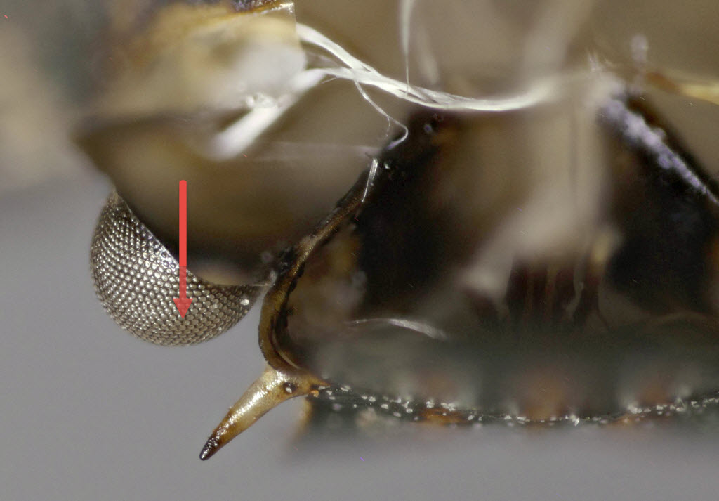
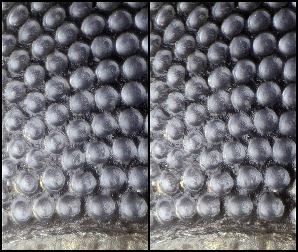
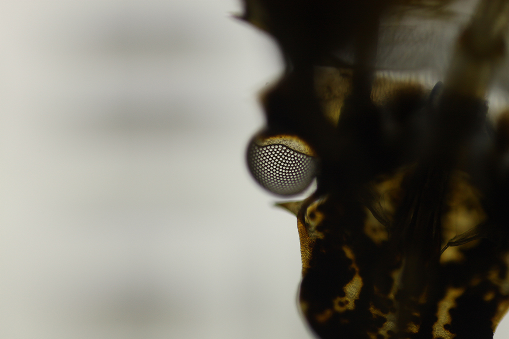
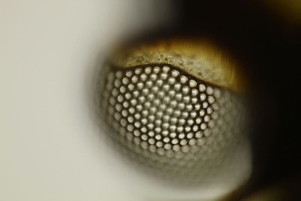
At lower left we see the ommatidial lenses themselves, shifted into focus by the overall curvature of the compound eye.
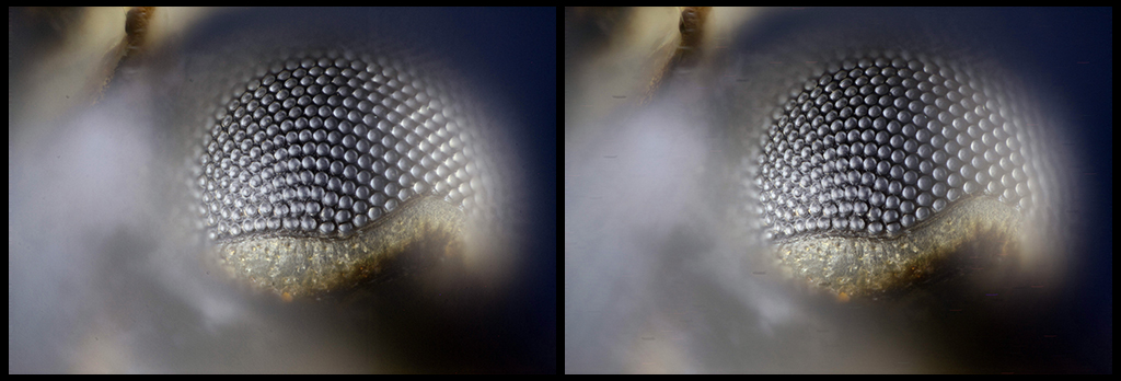
The above is 100% crop of a single image, Mitutoyo 20X M Plan Apo objective with Raynox DCR 150 tube lens, on Canon T1i sensor (pixel pitch 4.69 microns).
Following are some images that may help to make more sense of what's being shown.
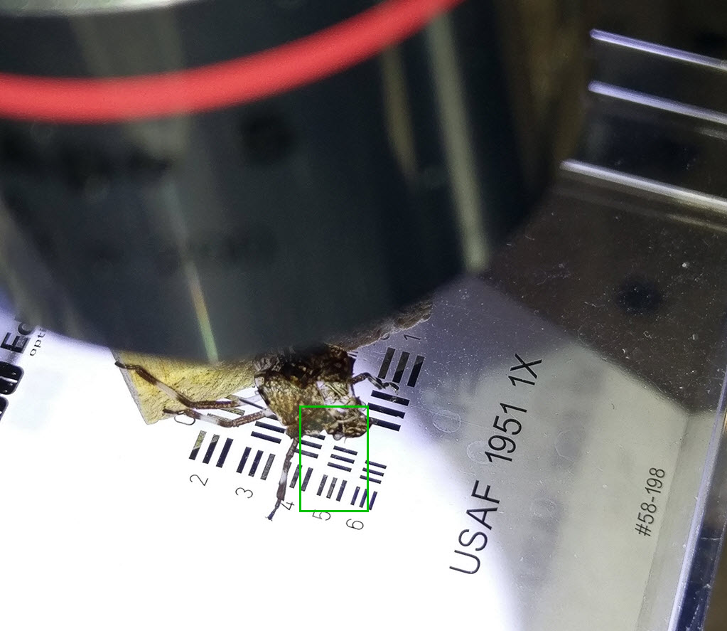
First, the overall setup. This is just a shed skin that happened to end up with the compound eye corneas oriented almost horizontally, positioned over a chrome-on-glass USAF resolution chart, illuminated from below by a small flashlight bounced off an index card. We're concerned with the stuff inside the green rectangle.
Here is the whole frame, as seen through a 5X objective, focused on the resolution chart.
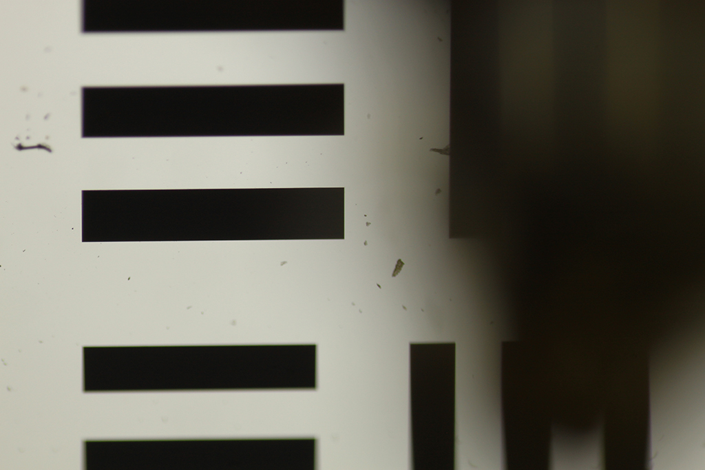
Again the whole frame at 5X, but now focused on the ommatidia images. This focal plane is 4.395 mm higher than the resolution chart.
Here, the whole frame at 20X. The first image shown in this thread is a 100% crop from this frame.
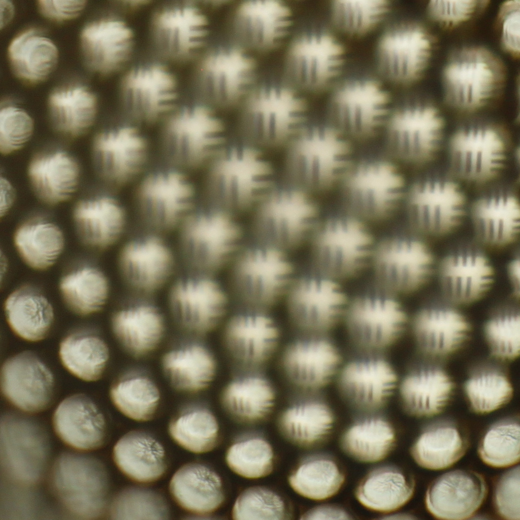
Comparing the resolution chart seen directly, versus through the ommatidia lenses, it's clear that we're looking at real inverted images formed behind the corneal lenses.
Comparing the size of the resolution chart bars as seen directly and through the ommatidia lenses, the magnification of the ommatidia lenses calculates out to be 1/113 X. Then from the 4.395 mm separation of focal planes and the magnification, the effective focal length of the ommatidia lenses calculates to be about 38 microns. The separation between adjacent ommatidia is about 27 microns, so allowing for the seams, these lenses are about f/1.8 .
I give thanks to Giorgio Pattarini (username patta) for our discussion in https://www.photomacrography.net/forum/ ... 27&t=42758, which prompted me to look again at this specimen. My earlier impression of it -- formed too quickly without looking closely -- was totally incorrect.
--Rik
Images formed by ommatidia lenses of BMSB shed cuticle
Moderators: rjlittlefield, ChrisR, Chris S., Pau
- rjlittlefield
- Site Admin
- Posts: 23624
- Joined: Tue Aug 01, 2006 8:34 am
- Location: Richland, Washington State, USA
- Contact:
Re: Images formed by ommatidia lenses of BMSB shed cuticle
So your getting an image through two layers of ommatidia, i.e. top and bottom of compound eye?
- rjlittlefield
- Site Admin
- Posts: 23624
- Joined: Tue Aug 01, 2006 8:34 am
- Location: Richland, Washington State, USA
- Contact:
Re: Images formed by ommatidia lenses of BMSB shed cuticle
No, just one layer.
Imagine the eye separating from the rest of the head everywhere except along its bottom edge. Then hinge the whole thing downward so that it ends up sort of like a bowl, open on the top.
The objective, looking down, is looking through a part of the eye that would have been lateral in life.
--Rik
Imagine the eye separating from the rest of the head everywhere except along its bottom edge. Then hinge the whole thing downward so that it ends up sort of like a bowl, open on the top.
The objective, looking down, is looking through a part of the eye that would have been lateral in life.
--Rik
- rjlittlefield
- Site Admin
- Posts: 23624
- Joined: Tue Aug 01, 2006 8:34 am
- Location: Richland, Washington State, USA
- Contact:
Re: Images formed by ommatidia lenses of BMSB shed cuticle
Here is an sideways view that I hope makes this more clear. This is looking from head to tail, perpendicular to optical axis. Red arrow represents the view of the objective.rjlittlefield wrote: ↑Fri Dec 11, 2020 12:17 amImagine the eye separating from the rest of the head everywhere except along its bottom edge. Then hinge the whole thing downward so that it ends up sort of like a bowl, open on the top.
The objective, looking down, is looking through a part of the eye that would have been lateral in life.
--Rik
Re: Images formed by ommatidia lenses of BMSB shed cuticle
This is fascinating! I can see the convex curvature of the exterior (distal) surface of the cuticle lens, but wonder about the interior surface contour. Is it spherical and continuous spanning the compound eye or dimpled by each ommatidium? Does it provide the mold to shape the corneal lenses revealed when the cuticle is discarded?
Cool stuf!
Keith
Cool stuf!
Keith
- rjlittlefield
- Site Admin
- Posts: 23624
- Joined: Tue Aug 01, 2006 8:34 am
- Location: Richland, Washington State, USA
- Contact:
Re: Images formed by ommatidia lenses of BMSB shed cuticle
I see it that the inside surface of each ommatidium is slightly convex toward the center of the eye. That is, each ommatidium lens is bi-convex, strong on the outside, weak on the inside. These are not forming a mold to shape the corneal lenses of the next instar.BugEZ wrote: ↑Sat Dec 12, 2020 7:53 amThis is fascinating! I can see the convex curvature of the exterior (distal) surface of the cuticle lens, but wonder about the interior surface contour. Is it spherical and continuous spanning the compound eye or dimpled by each ommatidium? Does it provide the mold to shape the corneal lenses revealed when the cuticle is discarded?
Here are a couple of crossed-eye stereo pairs of the interior of the cuticle. First one is the whole frame at 20.8X, second is a 100% crop, both on T1i sensor (22.3mm x 14.9mm, 4752 x 3168 pixels).
Illumination is from the side with a paper-diffused LED flashlight shining through the cornea on the right side of the specimen. So, the illumination for the ommatidia imaged sharply here has been diffused again, by passing through ommatidial lenses that are mostly seen in the whole-frame image as out-of-focus but creating veiling glare on the right side.
120 frames with step size 0.001 mm, stereo shift is plus and minus 5 degrees = +-0.998%, giving 10 degrees of stereo separation.
--Rik
Re: Images formed by ommatidia lenses of BMSB shed cuticle
Thanks Rik. The convex beads on the interior are amazing! In the transition between instars the vision must get pretty degraded.
Keith
Keith