Bdelloid rotifer, midge larva (polarized)

Images made through a microscope. All subject types.

Moderators: rjlittlefield, ChrisR, Chris S., Pau

Charles Krebs
Posts: 5865
Joined: Tue Aug 01, 2006 8:02 pm
Location: Issaquah, WA USA
Contact:

Bdelloid rotifer, midge larva (polarized)

Post by Charles Krebs »

Haven't done one of these rotifers in a while, so here it is. :wink:

The bdelloid rotifers were actually the reason I decided that I had to have electronic flash. I was so curious to see in better detail the movement pattern of the cilia.

The midge larva is an example of another lighting approach I like to use. By using crossed polarizers (one below the subject, one above... but usually not crossed to full "extinction") the muscle structure of many microscopic creatures can be easily seen. Normally these muscles are clear and colorless in regular brightfield illumination.

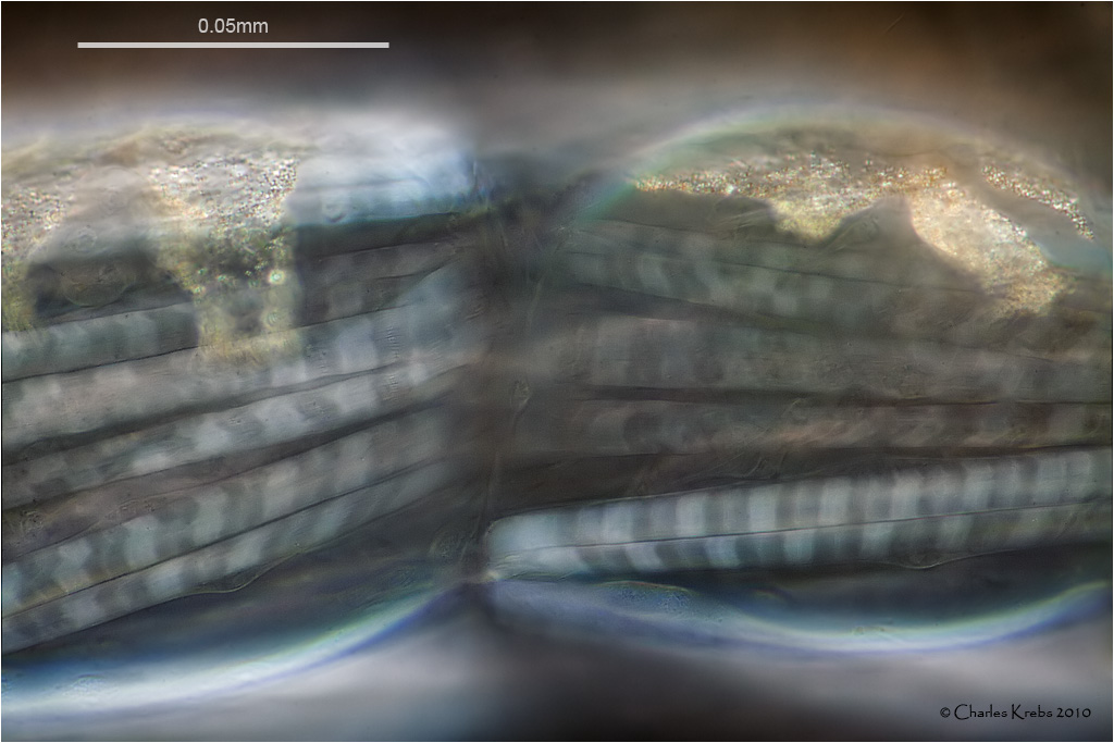
Olympus BHS. 40/0.95 S Plan Apo. 1.67 NFK. Canon 50D. DIC illumination.
Image


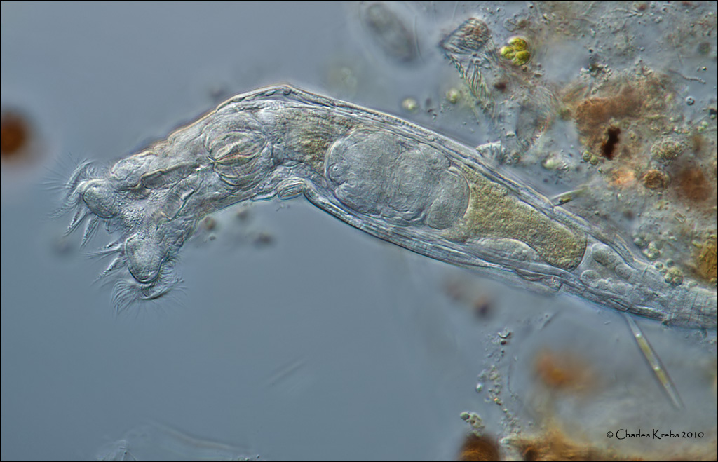
Olympus BHS. 10/0.40 S Plan Apo. 1.67 NFK. Canon 50D. Polarized brightfield illumination.
Image

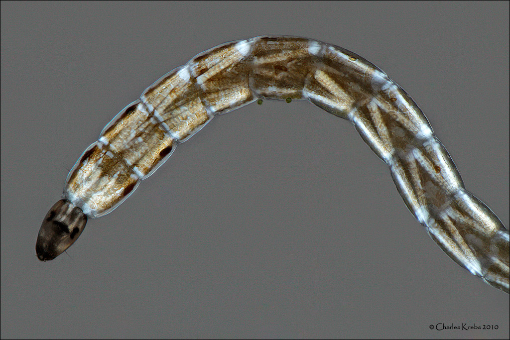
Olympus BHS. 60/1.40 S Plan Apo. 1.67 NFK. Canon 50D. Polarized brightfield illumination.
Image :wink:

rjlittlefield
Site Admin
Posts: 23625
Joined: Tue Aug 01, 2006 8:34 am
Location: Richland, Washington State, USA
Contact:

Post by rjlittlefield »

Magical!

--Rik

Pau
Site Admin
Posts: 6071
Joined: Wed Jan 20, 2010 8:57 am
Location: Valencia, Spain

Post by Pau »

Charles,
first and second are excellent but...
:shock: ...last one is absolutely extraordinary. I never saw a comparable light micrography of striated muscle whith all this detail and clarity in the bands, it's similar to low power TEM images shown in biology textbooks, but alive!:smt038 . This muscle cells must be really big.
Last edited by Pau on Mon Jun 14, 2010 2:02 pm, edited 1 time in total.
Pau

rjlittlefield
Site Admin
Posts: 23625
Joined: Tue Aug 01, 2006 8:34 am
Location: Richland, Washington State, USA
Contact:

Post by rjlittlefield »

I just noticed the "60/1.40". That image seems to have a lot of DOF. Single frame or stacked? If stacked, how deep and how did you keep the subject from moving?

--Rik

Charles Krebs
Posts: 5865
Joined: Tue Aug 01, 2006 8:02 pm
Location: Issaquah, WA USA
Contact:

Post by Charles Krebs »

Thanks for the kind remarks.

The third image is a single frame. The objective is a 1.40 but the condenser is 0.90. The midge is constrained on the "Z" axis by the cover slip (but not crushed). It could still "wriggle" on x/y axis. Electronic flash eliminates motion problems. BTW... the crossed polarizers really suck up the light. This was one of the very few times I needed to use my flash at full power (ISO 100 on camera). As is often the case, it took many images at different focus to eventually get the one where things lined up nicely in the same plane. Since these are essentially "clear", the focus is best set along the outer edge of the cylindrical muscles. Then the edges have some sharpness, and whatever is inside the muscle at that depth also has some sharpness. (An "optical section")

RogelioMoreno
Posts: 2979
Joined: Fri Nov 20, 2009 11:24 am
Location: Panama

Post by RogelioMoreno »

Charles,

Very nice!

Rogelio

morfa
Posts: 554
Joined: Mon Oct 12, 2009 2:14 pm
Location: Stockholm, Sweden
Contact:

Post by morfa »

Beautiful and inspiring! I very much enjoyed reading the discussion as well. I was surprised to learn that the third image was not stacked but considering it doesn't show surface detail but rather a cross section it makes sense!

JMak
Posts: 21
Joined: Mon Jul 26, 2010 5:38 pm
Location: United Kingdom

Post by JMak »

I am amazed. I didn't know it was possible to see muscles of such small creatures!

Cactusdave
Posts: 1631
Joined: Tue Jun 09, 2009 12:40 pm
Location: Bromley, Kent, UK

Post by Cactusdave »

Wonderful images of muscle. Superb.
Leitz Ortholux 1, Zeiss standard, Nikon Diaphot inverted, Canon photographic gear

Post Reply Previous topicNext topic