It is my first time photographing diatoms, and I had some trouble stacking them. Therefore, all these images are single shots. If anyone can shed some light on why I failed to stack these, I would love to know. Even with small stacks of 3 images, photoshop would misalign the dots on the frustules, what are some common pitfalls with diatoms? I suspect the main reason is there was too much other stuff in the water which it used to stack, I did not isolate these diatoms into clean water. Anyway, I think the single shots still turned out well, and diatoms are extraordinarily beautiful so little can go wrong.
I'm curious on people's thoughts about all the bacteria and other stuff present in the photo. Too distracting? Usually I like to see organisms in their natural environment, so I kinda like it. I did try cleaning up one of the images with a brush, though I did a whack job. What are people's thought on cleaning up images in general? Anyway, here are the photo's:
All shots are 100x oil DIC
 Img0234 by Mas Jansma, on Flickr
This species has a lovely way of attaching itself.
 Img0201 by Mas Jansma, on Flickr
 Img0187 by Mas Jansma, on Flickr
This is such a beautiful diatom, I wonder what that line down the middle is. Is it dividing?
 Img0142 by Mas Jansma, on Flickr
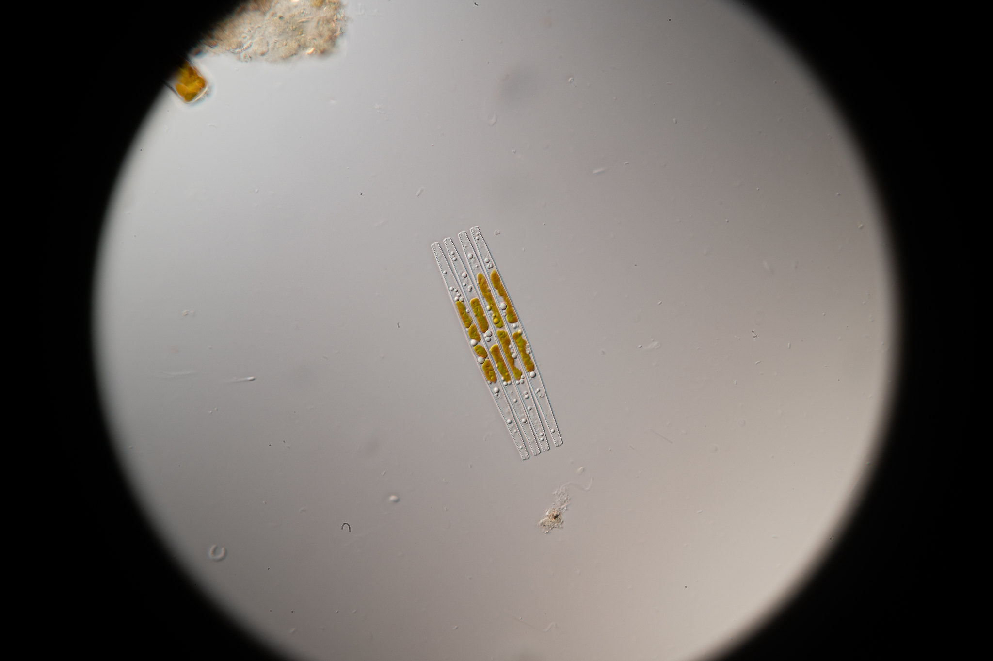 Img0133 by
 Mas Jansma, on Flickr
 Img0170 by Mas Jansma, on Flickr
 Img0164 by Mas Jansma, on Flickr
 Img0124 by Mas Jansma, on Flickr
You can see this nucleus dividing.
 Img0088 by Mas Jansma, on Flickr
I love the two perfectly shaped compartments on this individual. They were filled with tiny dots bouncing around. Some crystalline biocompound.
 Img0031 by Mas Jansma, on Flickr
The brushed up image:
 diatomee_shiny by Mas Jansma, on Flickr
I think using the brush did do something good in terms of pulling attention towards the subject. Maybe if I do a better job it will look less edited.
Hope you enjoyed these images. Let me know your thoughts.
Warm regards,
Mas


