
Coming back down to earth, most general photography is done by means of white light reflecting off of subjects, and recording that reflection. However, it is but one tiny way we can look at the world. Using different wavelengths (UV), polarized light, light wave interference (DIC), refracted light (darkfield), light wave retardation (lambda/quarter wave plates), and various combinations of these we can see so much more.
Here is the same amphipod specimen I found in my reef aquarium, but interrogated with most of the means to my disposal. I would have loved to have painted it by electron, but that is outside my reach (for now).
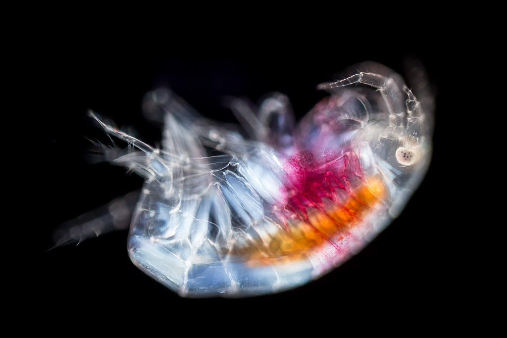
Amphipod by pwnell, on Flickr
Reef aquarium amphipod, 10x, DF+POL
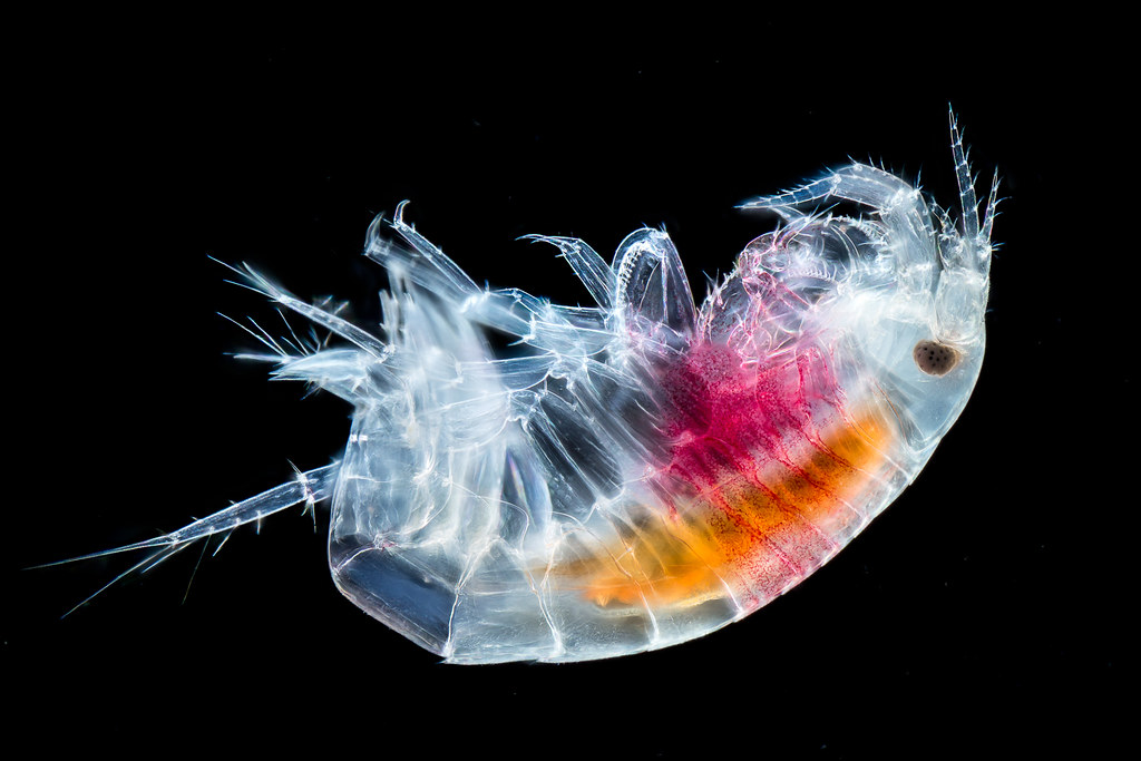
Amphipod by pwnell, on Flickr
Reef aquarium amphipod, 10x, DF+POL+LP, HF A+B
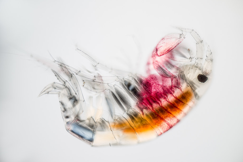
Amphipod by pwnell, on Flickr
Reef aquarium amphipod, 10x, POL+LP, HF B
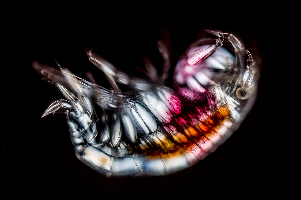
Amphipod by pwnell, on Flickr
Reef aquarium amphipod, 10x, POL
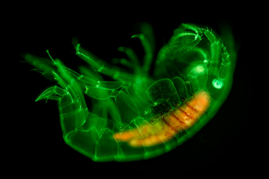
Amphipod by pwnell, on Flickr
Reef aquarium amphipod, 10x, FLUO-C4
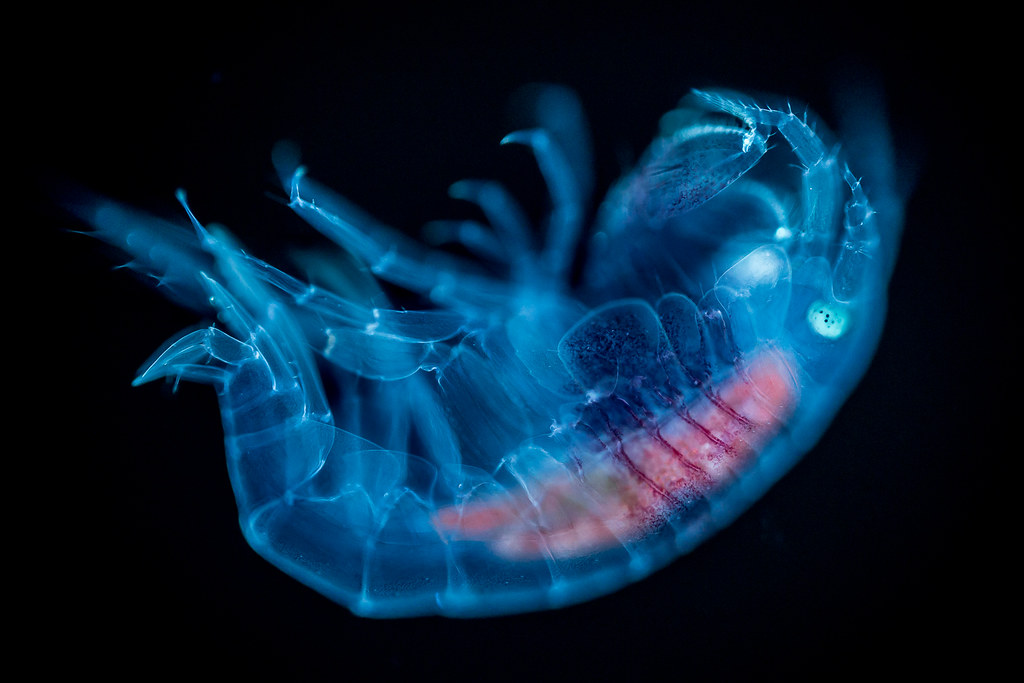
Amphipod by pwnell, on Flickr
Reef aquarium amphipod, 10x, FLUO-C6
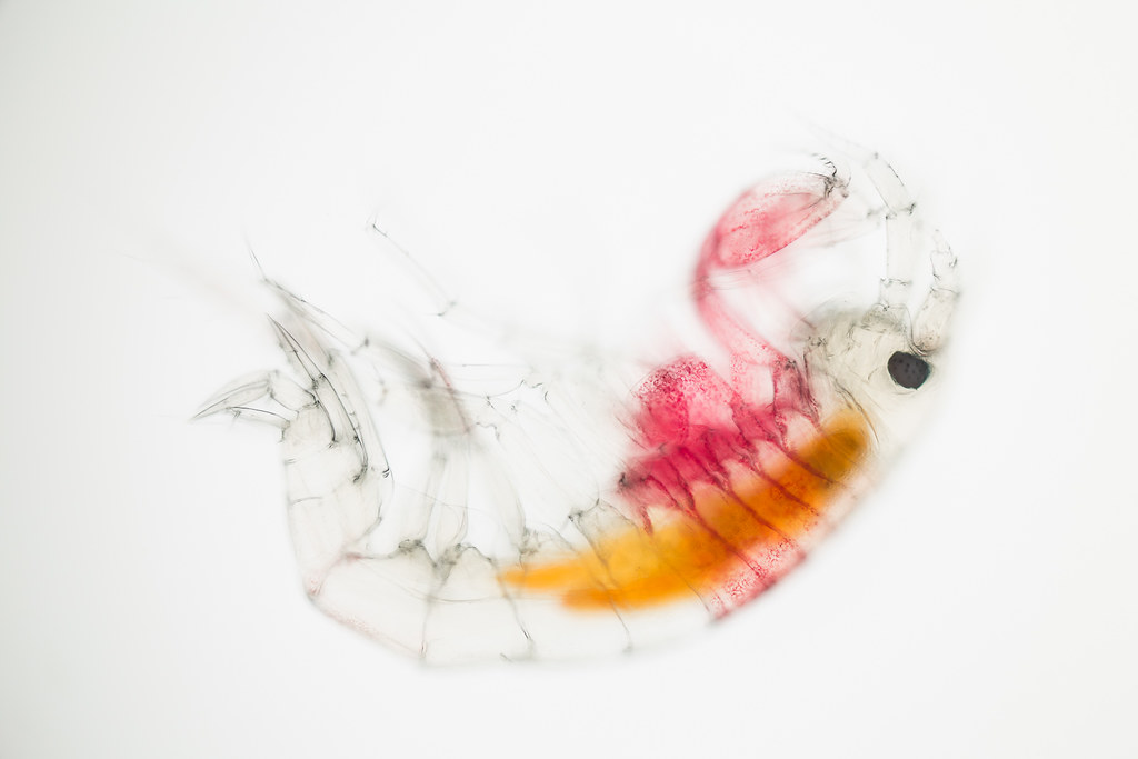
Amphipod by pwnell, on Flickr
Reef aquarium amphipod, 10x,BF
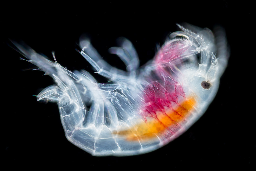
Amphipod by pwnell, on Flickr
Reef aquarium amphipod, 10x,DF
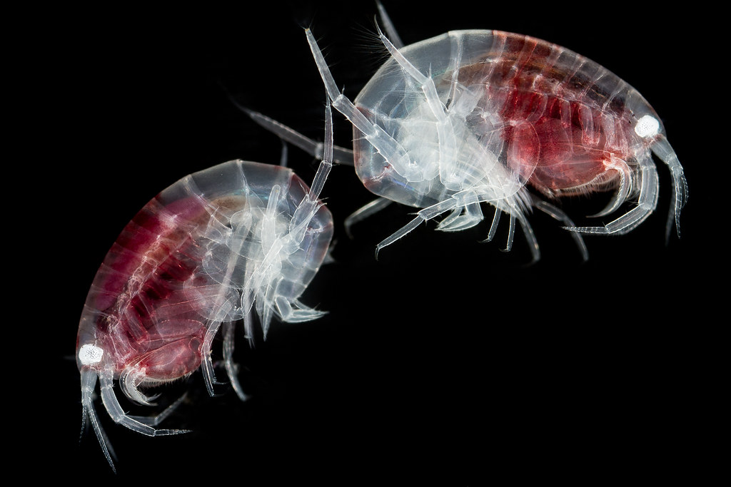
Amphipods by pwnell, on Flickr
Amphipods from Reef aquarium, Canon 1DX + MP-E65 @ 5:1, Reflected Brightfield (not microscope).


