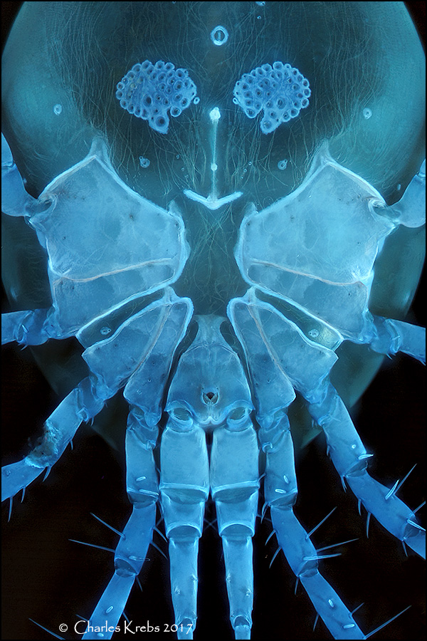
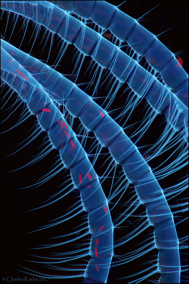
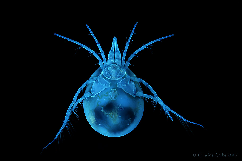
The last three were inspired by Jacek's wonderful water mites. These are not as exotic looking as some of the ones he has posted, but they sure do auto-fluoresce nicely under UV light.
50/0.50 Olympus LMPLFLN
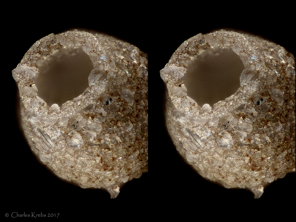
cross-eyed stereo 20/0.40 Olympus LMPLFLN
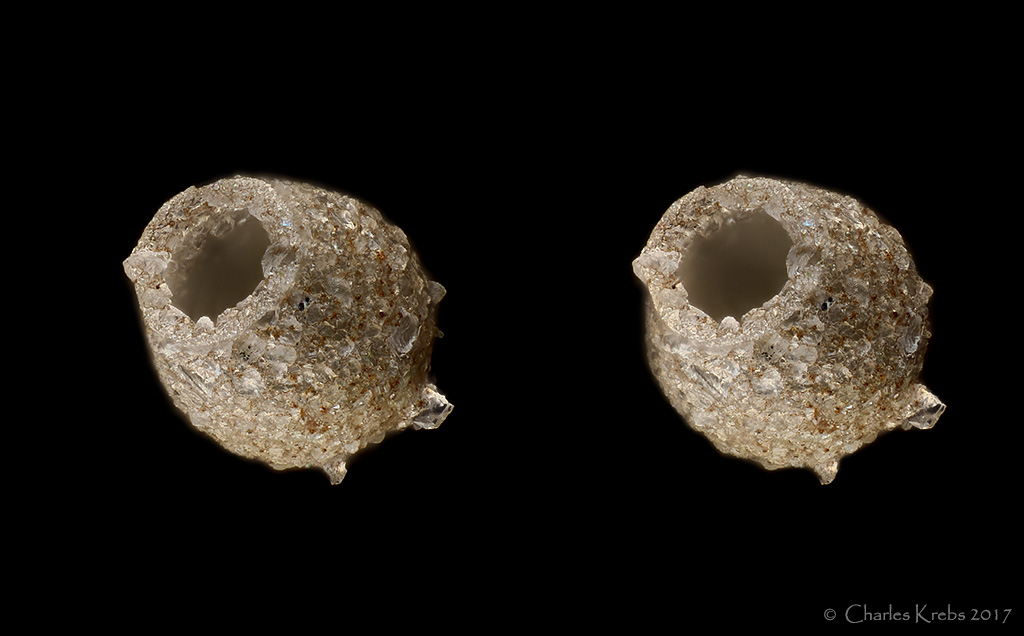
cross-eyed stereo 50/0.50 Olympus LMPLFLN
4/0.13 Olympus UPLFLN, 365nm UV excitation.
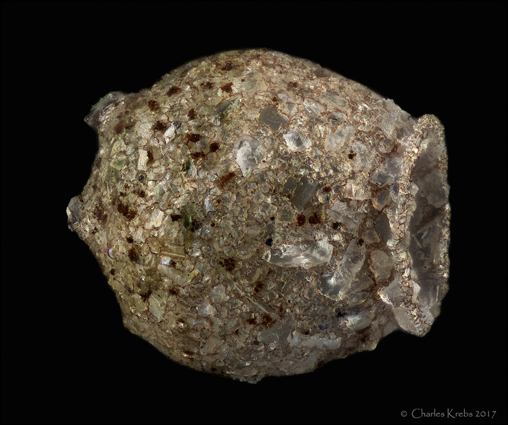
10/0.30 Olympus MPLFLN
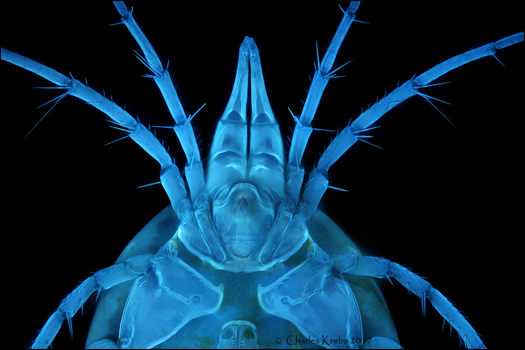
10/0.30 Olympus MPLFLN