Amoeba test and fluorescent water mite

Images made through a microscope. All subject types.

Moderators: rjlittlefield, ChrisR, Chris S., Pau

Charles Krebs
Posts: 5865
Joined: Tue Aug 01, 2006 8:02 pm
Location: Issaquah, WA USA
Contact:

Amoeba test and fluorescent water mite

Post by Charles Krebs »

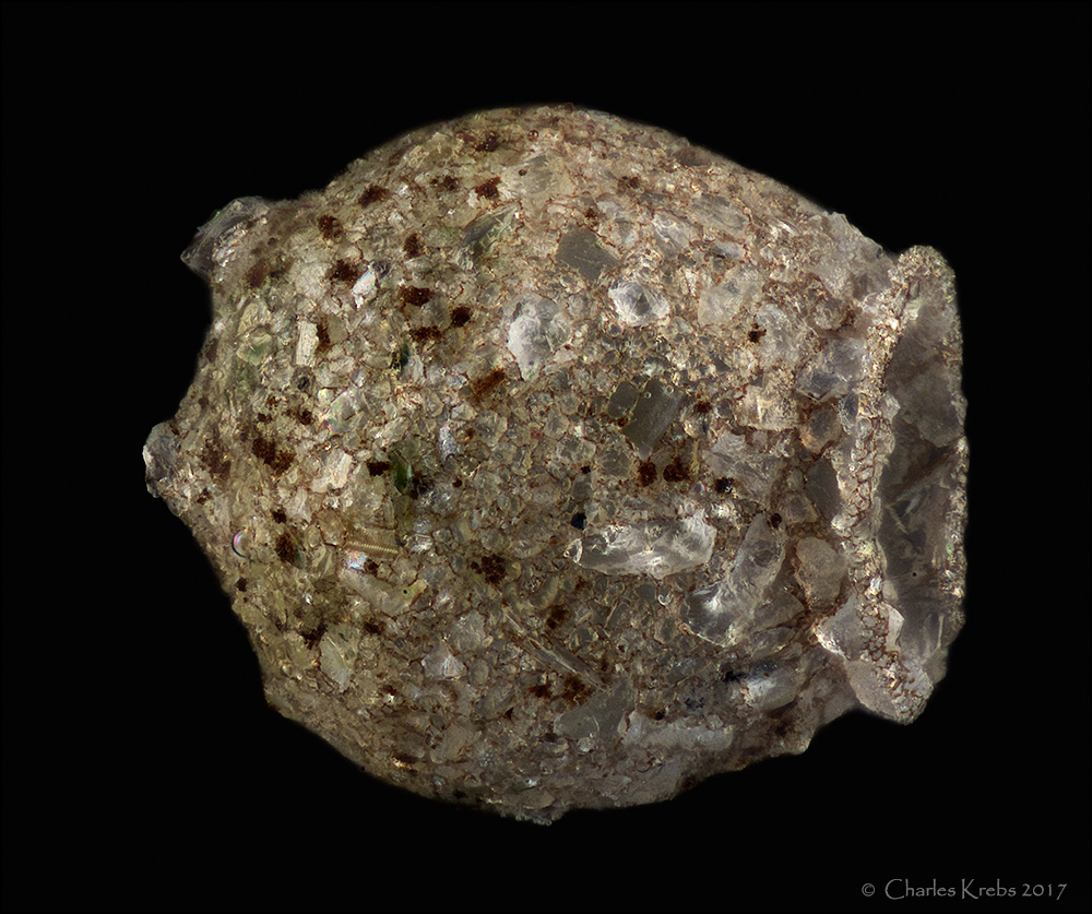
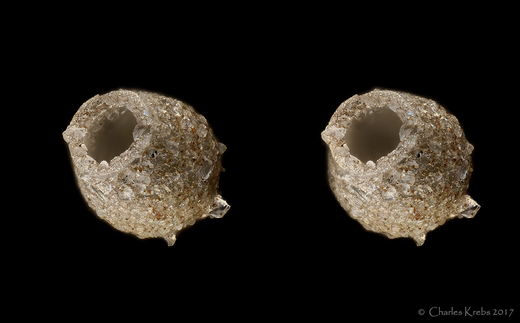
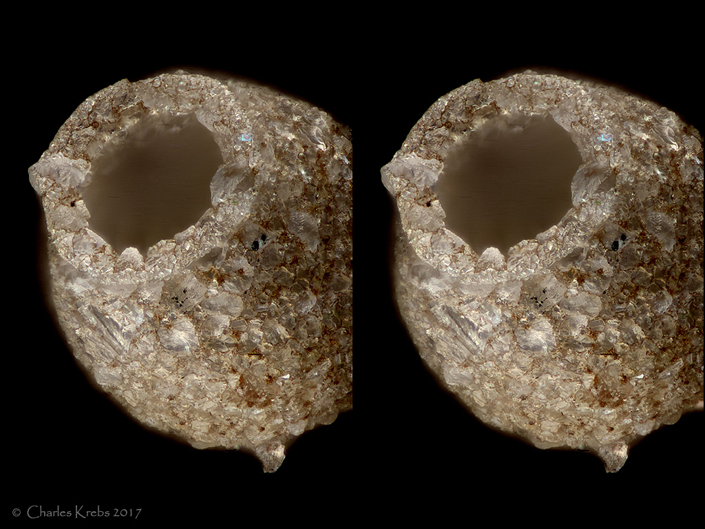
Here are a few subjects from a new "sample". Since I stated observing through a microscope, amoeba tests are a subject that always amaze me... perfect 'micro-pottery" made by a single celled amoeba!

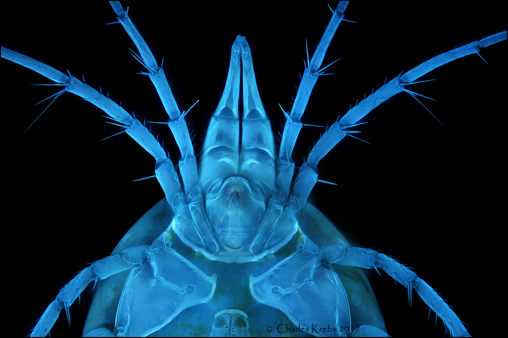
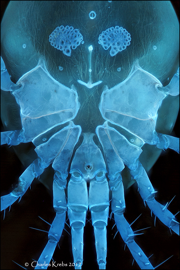
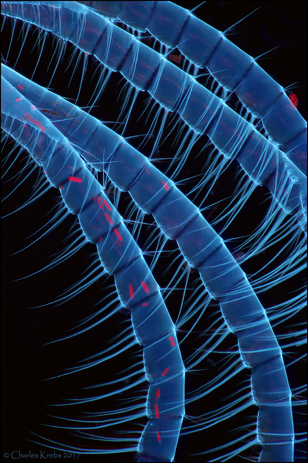
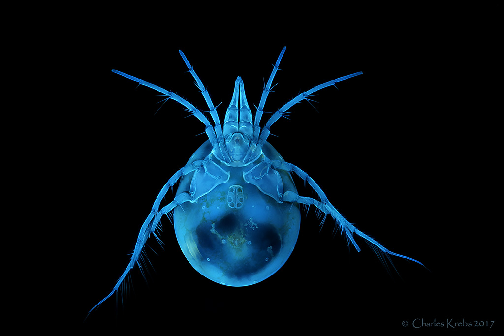
The last three were inspired by Jacek's wonderful water mites. These are not as exotic looking as some of the ones he has posted, but they sure do auto-fluoresce nicely under UV light.

50/0.50 Olympus LMPLFLN
Image

cross-eyed stereo 20/0.40 Olympus LMPLFLN
Image

cross-eyed stereo 50/0.50 Olympus LMPLFLN
Image

4/0.13 Olympus UPLFLN, 365nm UV excitation.
Image

10/0.30 Olympus MPLFLN
Image

10/0.30 Olympus MPLFLN
Image

zzffnn
Posts: 1896
Joined: Thu May 22, 2014 1:25 pm
Location: Houston, Texas, USA
Contact:

Post by zzffnn »

Great images, Charles!

Your seemed to have changed positions of the test a bit for framing, how did you do it? Not with a micromanipulator, I assumed?

Thank you for sharing!
Selling my Canon FD 200mm F/2.8 lens

Charles Krebs
Posts: 5865
Joined: Tue Aug 01, 2006 8:02 pm
Location: Issaquah, WA USA
Contact:

Post by Charles Krebs »

zzffnn wrote: Your seemed to have changed positions of the test a bit for framing, how did you do it? Not with a micromanipulator, I assumed?
There are two different specimens represented here. I placed about 7 of these tests (same species) on a slide and let the water evaporate. Just before all the water was completely gone I very carefully (under a stereo scope) used a fine needle to stand a few of them on end, so that the test aperture was pointed upward. Once dry enough to photograph I don't try to move them. It's possible, but they really are quite fragile.

Lou Jost
Posts: 5987
Joined: Fri Sep 04, 2015 7:03 am
Location: Ecuador
Contact:

Post by Lou Jost »

Amoeba tests blow my mind. The little rock fragments look like they've been fitted together as precisely as the finest Inca stonework-- yet without the use of tools, or eyes, or hands, or a brain.

How does one find them? I'd love to see a real one.

zzffnn
Posts: 1896
Joined: Thu May 22, 2014 1:25 pm
Location: Houston, Texas, USA
Contact:

Post by zzffnn »

Lou Jost wrote:How does one find them? I'd love to see a real one.
Lou,
You should be able to find them in many fresh water ponds of any size. They are slow bottom feeders and should stick to the bottom of your container. To be sure, ask http://www.arcella.nl/contact
Selling my Canon FD 200mm F/2.8 lens

Lou Jost
Posts: 5987
Joined: Fri Sep 04, 2015 7:03 am
Location: Ecuador
Contact:

Post by Lou Jost »

Thank you Fan for the link to that treasure-trove of information! Everything I could ever want to know about them.

Marek Mis
Posts: 2586
Joined: Sat Jul 10, 2010 9:56 am
Location: Suwalki, Poland
Contact:

Post by Marek Mis »

Super !!

Marek

Jacek
Posts: 5357
Joined: Sun Oct 02, 2011 7:00 am
Location: Poland

Post by Jacek »

Your photos are always perfect. I'm your fan.

Water mite, has it been the same, or did you help him, how did you "sleep"?

Your UV light will pass a lot of blue, it's not pure 365 nm.
First Water mite is Limnesia koenikei she does not shine all, no armor.
Use the excite filter to see the differences.

I'm sorry for my photo in your post, but it's best to explain what I mean
Image

Charles Krebs
Posts: 5865
Joined: Tue Aug 01, 2006 8:02 pm
Location: Issaquah, WA USA
Contact:

Post by Charles Krebs »

Jacek wrote:Your UV light will pass a lot of blue, it's not pure 365 nm.

Jacek, what is the wavelength range of the excitation filter you used? I realize these lights have some "spillover" wavelengths longer than 365nm. For that reason I use 310-385nm excitation filters on both of the lights I use for photography. There should be no violet or blue light illuminating the subject (nothing longer than 385nm). These mites fluoresce as seen above... very much like the shed barnacle cuticle below:
Image

Jacek
Posts: 5357
Joined: Sun Oct 02, 2011 7:00 am
Location: Poland

Post by Jacek »

I understand, you are probably the stronger source of light from me.

Write how you slept water mite ?

carlos.uruguay
Posts: 5358
Joined: Thu Feb 23, 2012 8:05 pm
Location: Uruguay - Montevideo - America del Sur
Contact:

Post by carlos.uruguay »

Perfect and beautiful photos!! as usual
The shell interior in the second and third pair of photos have digitally retouched?

Smokedaddy
Posts: 1965
Joined: Sat Oct 07, 2006 10:16 am
Location: Bigfork, Montana
Contact:

Post by Smokedaddy »

Amazing ... Charles where do you find amoeba's like that? To be honest, I didn't even know they existed. I do a LOT of 4-wheeling and go where very view people go. I've collected small water samples a couple of time where I've seen green moss growing in a couple of streams but haven't seen anything at all yet. I was actually looking for diatoms.

Olympusman
Posts: 5090
Joined: Sun Jan 15, 2012 12:31 pm

Water mite

Post by Olympusman »

Loved the whole body watermites. Loved the details as well.

Mike
Michael Reese Much FRMS EMS Bethlehem, Pennsylvania, USA

Charles Krebs
Posts: 5865
Joined: Tue Aug 01, 2006 8:02 pm
Location: Issaquah, WA USA
Contact:

Post by Charles Krebs »

Lou Jost wrote:How does one find them? I'd love to see a real one.
Smokedaddy wrote:Amazing ... Charles where do you find amoeba's like that? To be honest, I didn't even know they existed.
You can usually find them in most fresh water ponds/bogs/streams/lakes where you would typically get samples. Be sure to collect some of the debris, vegetation, and dirt at the bottom of the water where you are sampling. I also find them in the dirt at the base of a moss sample at the edge of streams. While you can certainly find them on sample vegetation I generally have the best luck looking through the debris that settles out at the bottom of a jar or sample container. The larger heavier ones will often settle to the bottom and if you draw off the silty layer you can find them down there. I think the trickiest part is "training" your eye what to look for. A 20X to 30X stereo view seems about right for me when picking through a sample that has potential. Once you find a few I think you will be surprised at how common they really are.

If you find them interesting you should look through the plates of this old book by Joseph Leidy (published in 1879). Every time I look through these I am amazed by the work he did.

https://user.xmission.com/~psneeley/Personal/FwrPLA.htm

For more about Leidy and this book see here:
https://user.xmission.com/~psneeley/Personal/Leidy.htm
carlos.uruguay wrote:The shell interior in the second and third pair of photos have digitally retouched?
No, not at all. I was also surprised about the appearance of the interior. I expected to see more of the interior surface. But there were amoeba inside so perhaps that explains it. There was very little retouching at all on the two stereo shots besides cleaning up a small amount of outside "halo". If I find that a shot needs a lot of "clean-up" after stacking I usually don't bother with a stereo version since it is very time consuming and (for me at least) difficult to do without being quite obvious in the final result.

WalterD
Posts: 616
Joined: Mon Jul 06, 2015 12:01 pm
Location: Rotterdam, the Netherlands
Contact:

Post by WalterD »

Lovely set ; the amoeba are just spectacular!
I can imagine they're difficult to spot in a sample.

Thanks for sharing.


:D

Post Reply Previous topicNext topic